Elbow dysplasia
What is elbow dysplasia? The term “dysplasia” means abnormal development of a tissue or organ. Elbow dysplasia therefore means that there has been abnormal development of the elbow joint.
The consequence of this abnormal development is that the three bones of the joint (the humerus, radius and ulna) do not fit together perfectly leading to areas of abnormally high contact pressure. This in turn leads to one of a number of different problems (more than one of which may occur in the same joint at the same time):
- Fragmented medial coronoid process (FCP)
- Osteochondritis dissecans (OCD)
- Ununited anconeal process (UAP)
- Medial compartment disease
“Elbow dysplasia” is really an umbrella term for a number of different conditions of this joint. In most cases both elbows are affected and dogs are much more commonly affected than cats.
What are the signs of elbow dysplasia?
Regardless of which of the above four conditions is present, the signs of elbow dysplasia are the same. Typically affected dogs show lameness of one or both front legs, stiffness (especially after lying down), and reluctance to exercise. Often the feet of the front limbs appear turned out. Coming down stairs is often awkward as the elbows are sore. The most common age when the signs are first seen is between 6 and 10 months of age, but some dogs only present as middle aged or older adults when they have advanced arthritis.
In most cases, orthopaedic examination reveals elbow swelling and pain, with a restricted range of movement as the joint becomes thickened as a result of arthritis. In a small number of dogs the elbows are painful but not swollen and the diagnosis in these cases can be challenging.
When is ED required for your dog?
If you suspect that your dog is suffering from ED, it is important to have your animal examined on time, as the condition not only causes pain, deformation and lameness, but also long-term wear of osteoarthritis in your dog.
How is the diagnosis confirmed?
Elbow dysplasia is a collective name for five different disorders: LPC, LPA, OCD, incongruence and flexorenthesopathy. To make the correct diagnosis, the vet will examine not only the painful leg but also the other three legs of your dog. This way you can see if there are other orthopedic problems.
The vet will usually take x-rays of your dog’s elbows. When taking photos, it is important that your dog’s legs are straight. Taking an X-ray is a matter of seconds, so it is usually not very stressful for your dog and anesthesia is not necessary. Only if your dog cannot stand still because of the pain or if he is fiercely resisting, will he get a he slight anesthetic
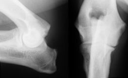
- Fragmented medial coronoid process (FCP)
The radiographs here are from a 10 month old labrador retriever with mild arthritis. Whilst the changes are not specific to elbow dysplasia, this dog actually has a fragmented coronoid process.
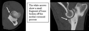
The CT scan images here confirm that the dog actually has a fragmented medial coronoid process.
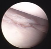
An arthroscopic view of the inside of this dog’s joint is shown below. The needle on the right side of the image is lifting up a fragment that has broken away from the medial coronoid process.
Surgical removal of these fragments of bone leads to a good improvement in 60-70% of dogs but not all dogs respond to treatment. We recommend arthroscopic surgery rather than open surgery as not only is it the best way to inspect the joint, but it is also minimally invasive and both elbows can be operated on under the same anaesthetic if required.
The fact that the bones of the joint do not fit perfectly together means that arthritis will develop in all cases as the dogs get older. If there is a step between the joint surfaces of the radius and ulna then it may be necessary to cut the ulna (“ulnar osteotomy”) to improve the alignment of the bones and minimise the on-going cartilage wear.
- Osteochondritis dissecans (OCD)
In this condition an area of cartilage on the bottom of the humerus (the “humeral condyle”) does not develop normally and lifts off. This is also associated with abnormal bone development underneath the area of affected cartilage.
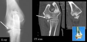
These images (x-ray on the left, CT scan on the right) are of an 8month old rottweiler with OCD. The area of abnormal bone development is shown by the arrow.
Removal of the cartilage flap improves the lameness in many cases but like FCP not all dogs respond to treatment. The cartilage does not grow back again, leaving a gap in the normal joint surface. Consequently arthritis develops in all cases. Once again, we would recommend arthroscopic joint inspection as some dogs have both FCP and OCD. The cartilage flap can be readily removed under (minimally invasive) arthroscopic guidance.
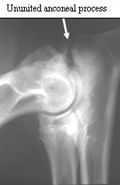
- Ununited anconeal process (UAP)
The anconeal process of the ulna is part of the elbow joint. This part of the bone develops from a separate centre of growth to the rest of the ulna but the two parts join together at around 4 months of age. Abnormal development of the elbow (dysplasia) can lead to pressure on the anconeal process which stops it from joining on to the rest of the ulna. The x-ray here shows a dog with an ununited anconeal process.
In most dogs a CT scan is not necessary to make this diagnosis as the ununited anconeal process shows up well on an x-ray.
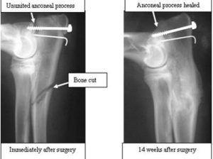
If the diagnosis is made in young dogs (generally under 8 months of age), then the ununited anconeal process can be fixed back on with a screw. In these cases the ulna needs to be sectioned (“ulnar osteotomy”) to take the abnormal pressure off the anconeal process and to allow it to heal. Arthroscopic examination of the joint is recommended as many dogs with ununited anconeal process also have other changes such as FCP. In older dogs the ununited anconeal process rarely joins back on to the ulna even with surgery and may need to be removed. In dogs with mild lameness it is usually best to leave it in place.
The x-rays here are from a dog that has had surgery for UAP. A screw and a pin have been used to secure the loose anconeal process and the ulna has been cut. The x-ray on the right shows the anconeal process fused on to the ulna and healed bone cut 14 weeks later.
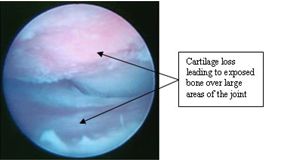
- Medial compartment disease (MCD)
In some dogs the abnormal pressure of one joint surface on the other leads to the cartilage being worn away. The underlying bone is then exposed and the joints become very inflamed and arthritic. This form of elbow dysplasia carries the poorest prognosis as it is not possible to reverse the cartilage loss. Several surgical procedures have been developed to try and move the forces of weight bearing away from these areas (proximal dynamic ulnar osteotomy and sliding humeral osteotomy). These have not been scientifically proven to be of clinical benefit and are a last resort.
Is surgery always recommended for elbow dysplasia?
In short, no.
The underlying problem in elbow dysplasia is abnormal development of the joint, and it is not possible to reverse the process and make the joint normal. All dogs with elbow dysplasia will develop arthritis to some degree even if they undergo surgery. This must be taken into consideration when deciding on whether to operate or not.
Many dogs can be managed effectively using non-surgical or “conservative” measures:
- Very careful control of body weight.
- Controlled exercise, avoiding boisterous activities such as running, turning at speed, chasing a ball, rough and tumble with other dogs, braking sharply and jumping down to land on the front legs.
- Hydrotherapy is beneficial as it works the muscles without overload the elbow joint. This is a good way to keep dogs fit and to help with weight control.
- Dietary supplements such as omega-3-fatty acids, glucosamine and chondroitin sulphate may relieve some of the joint discomfort and stiffness. Prescription diets are available for arthritic dogs and these can be beneficial.
- Prescription medication such as anti-inflammatory drugs may be required on either a daily or as-needed basis.
Preventing elbow dysplasia:
Preventing elbow dysplasia starts with choosing a puppy born to parents who have been checked and found free for elbow dysplasia. In addition, metered movement is very important. The same exercise is better every day than walking a day a week and very little the other days. The duration of the walk must also be adjusted to the age of the dog.
Nutrition
When feeding, we must keep a close eye on the calcium-phosphorus ratio and the absolute amount of calcium. Care must also be taken that not too much food is given so that your dog does not become overweight. It is important to discuss the level of exercise and nutrition with an expert, the breeder of your dog or your veterinarian.